
Overview
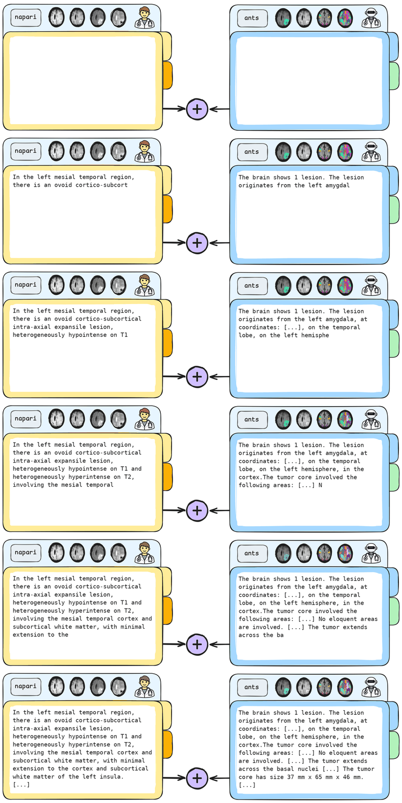
ReportX is a clinician-curated brain tumor MRI report dataset aligned with the BraTS-GLI-2023 dataset and published in the MICCAI 2026 paper ReportX: The BraTS Clinical Report Dataset. It includes 1,251 automatically generated reports for the full BraTS-GLI-2023 dataset, obtained by algorithms that use segmentation masks, atlas-based localization, and geometric measurements, together with 257 clinician-curated reports written by expert neuroradiologists. The official repository is available on GitHub.

Dataset Description
|
Field |
Value |
|---|---|
|
Source Dataset |
BraTS-GLI-2023 |
|
Annotation types |
Clinicians-Curated (CC) + Automatically-Derived (AD) |
|
Reports (CC+AD) |
257 |
|
Reports (AD) |
1,251 |
|
Atlas Space |
SRI24 |
|
Anatomical atlas |
236 anatom. regions |
|
Eloquent-area atlases |
4 areas: motor, speech-motor vision, speech-receptive |
|
License |
ReportX is a structured brain tumor MRI report dataset built on BraTS-GLI-2023. It provides 1,251 Automatically Derived reports (AD) for the full BraTS-GLI-2023 cohort, generated from tumor masks, atlas-based localization, and geometric measurements, together with a 257-case Clinician-Curated subset (CC) annotated by expert neuroradiologists while reviewing the four BraTS MRI modalities. The clinician-written fields describe tumor appearance, subregions, and contextual findings, while the automatically derived fields describe lesion count, tumor origin, laterality, anatomical involvement, eloquent-area overlap, size, edema extent, and other region-aware quantitative descriptors.
To support automatic localization, ReportX also releases the atlases used in the pipeline. These include a clinician-curated anatomical atlas, based on Parc116 and expanded with fully segmented subcortical and deep white-matter regions, together with four binary eloquent-area atlases for motor, speech-motor, speech-receptive, and vision functions. The atlases are defined in SRI24 space and are warped to each subject using ANTs-based deformable registration, enabling automatic identification of tumor origin, involved anatomical regions, and overlap with eloquent areas.
Dataset Structure
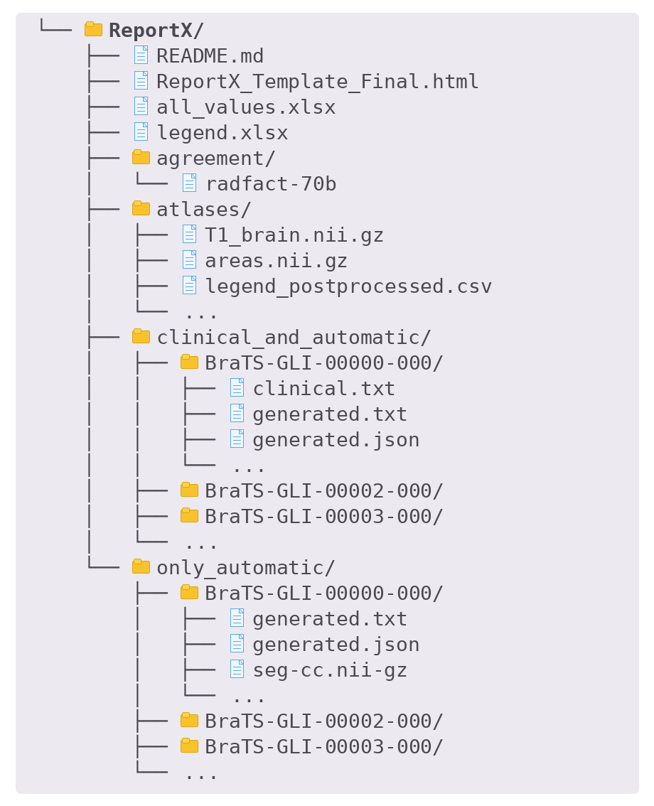
The downloadable ReportX package is organized into case-level folders and shared metadata resources. The clinical_and_automatic/ directory contains the 257 clinician-curated subset, with one folder per case. Each case includes the clinician-curated report clinical.txt, the automatically derived report generated.txt, a structured JSON with the automatic descriptors generated.json, the concatenated report concat.txt, and NIfTI files of tumor masks, warped anatomical atlases, and eloquent-area masks. The only_automatic/ directory follows the same structure but contains the 1,251 automatically derived reports and masks, without clinician-written text.
The atlases/ directory provides the reference anatomical and eloquent-area atlases used by the automatic pipeline, while legend.xlsx, legend_postprocessed.csv, and all_values.xlsx describe the anatomical labels, macroareas, depths, and sides. The splits/ directory contains the case split spreadsheets, and agreement/ includes the RadFact prompt used for factual-consistency evaluation. For full details, see the README included in the download.
Dataset Validation
ReportX was validated using a structured protocol that assesses both the reliability and the clinical completeness of the reports. The clinician-written fields were evaluated through inter-clinician agreement on 50 overlapping cases using RadFact, showing strong consistency between annotations from different radiologists. Report quality was further assessed with the Concept Coverage Score, which measures how well each report covers standardized neuro-oncological concepts from RadLex Playbook and VASARI MRI Feature Set, and Template Adherence, which evaluates the presence of clinically relevant reporting elements such as localization, size, morphology, necrosis, infiltration, perfusion, and diffusion.
The automatically derived fields were also validated on the same 50 cases by comparing the pipeline outputs with ad-hoc clinician annotations, measuring the accuracy of lesion count, tumor origin, origin side, anatomical areas involved by the tumor core and edema, and overlap with eloquent areas.
| Field | Precision | Recall | F1 Score | Prevalence |
| Areas (TC) | 73.90 | 96.84 | 83.83 | 6.25 |
| Eloq. areas (TC) | 80.64 | 99.02 | 88.89 | 0.13 |
| Areas (ED) | 84.05 | 99.70 | 91.21 | 12.69 |
| Field | Accuracy |
| Number of lesions | 98.00 |
| Origin location | 94.23 |
| Origin side | 98.36 |