IOP-Compass
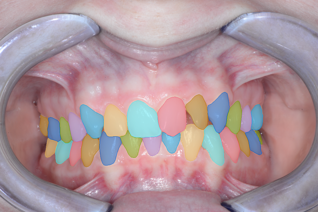
5,000 standardized intraoral photographs from 1,000 patients, with expert-verified FDI tooth-instance masks.
Overview
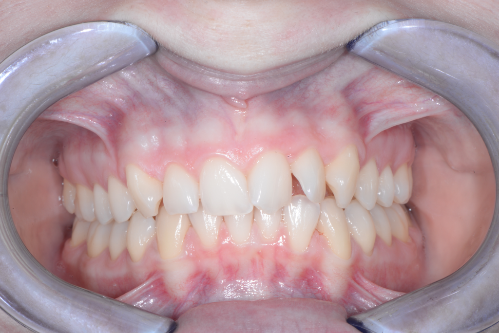
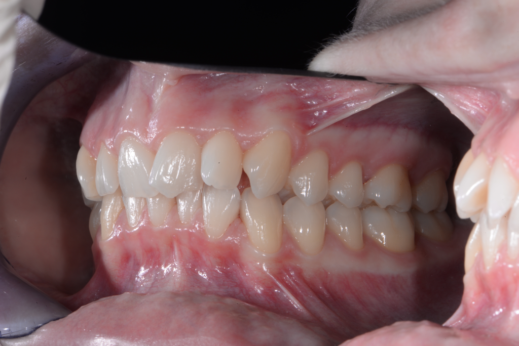
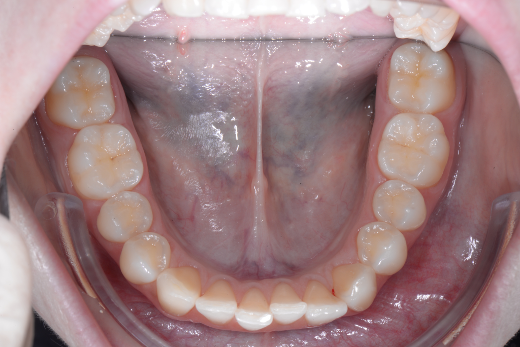
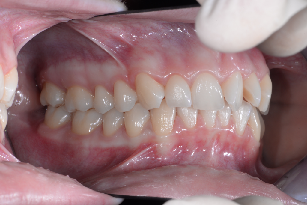
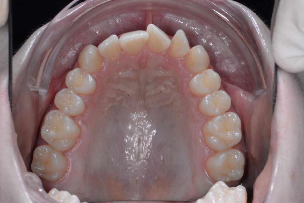
IOP-Compass is a comprehensive dataset of intraoral photographs from 1,000 patients, featuring 5,000 images with expert-verified tooth-instance masks labeled with FDI numbers. Each patient contributes five standardized orthodontic views, facilitating research in multi-view analysis and tooth segmentation. The images are sourced from the Bite2Text dataset and therefore follow the same image acquisition protocol, including the standardized intraoral views and capture conditions used in the original dataset.
Dataset Focus
The IOP-Compass dataset is designed to support research in tooth segmentation and multi-view analysis. Segmenting teeth in 2D intraoral photographs is a crucial step that can enhance the performance of more sophisticated algorithms, such as those used in orthodontic diagnosis and treatment planning.


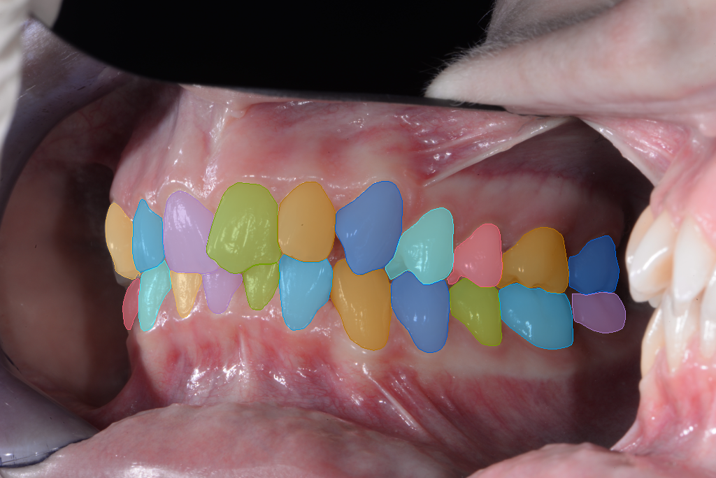
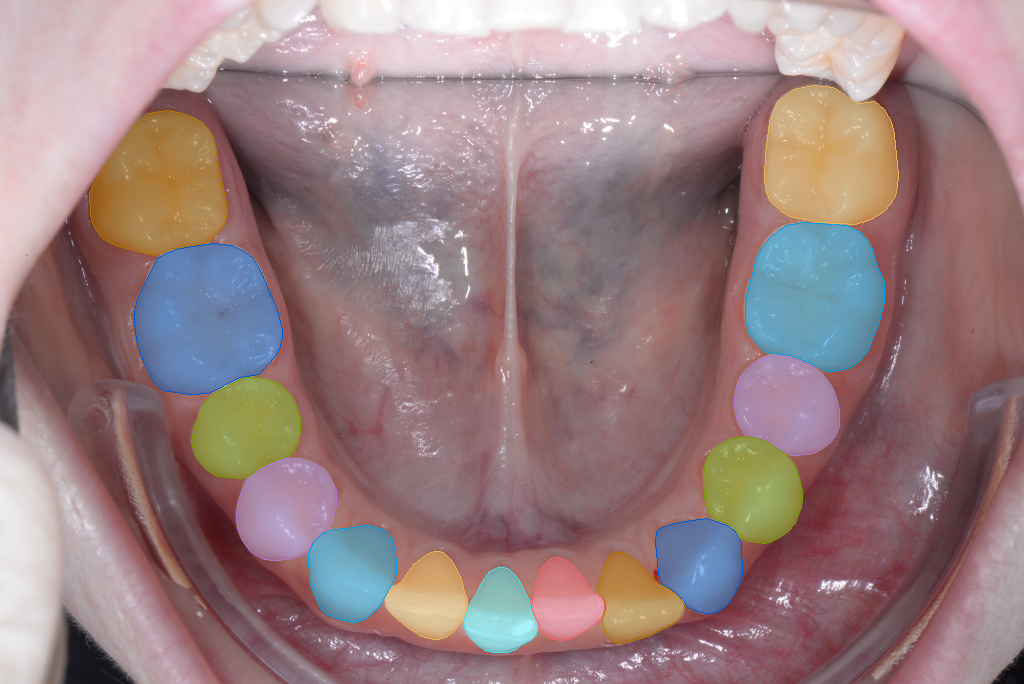
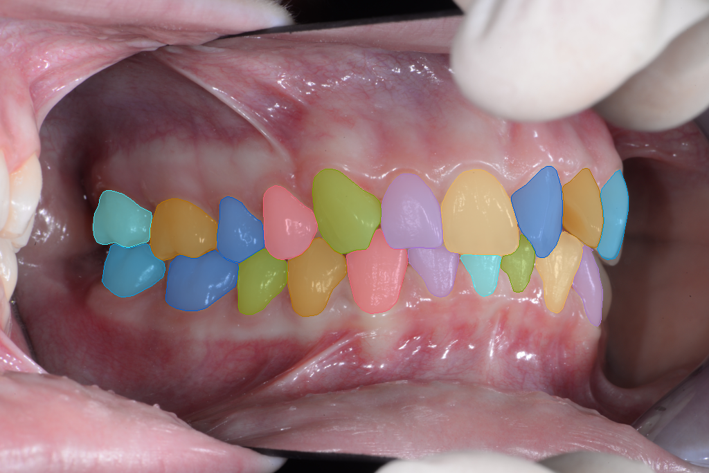
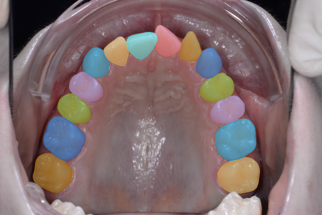
Structure & Features
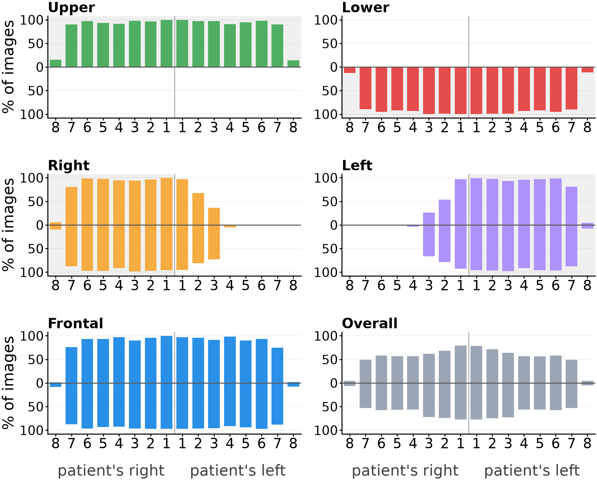
The images in the IOP-Compass dataset were collected from two different labs, both employing similar camera equipment and capture protocols. The use of a typical clinical mirror for intraoral photographs (IOPs) was standard practice, ensuring consistency across the dataset. This setup allows for reliable segmentation and analysis of tooth instances across orthodontic views.
Each case/patient is organized as:
IOP_Center_1.png: center raw IOP 2D RGB image;IOP_Center_1.json: overlay boxes with FDI;IOP_Upper_1.png: upper raw IOP 2D RGB image;IOP_Upper_1.json: overlay boxes with FDI;...